
UCSD Musculoskeletal Radiology
bonepit.com
Case of the week 003
|
|
UCSD Musculoskeletal Radiology bonepit.com Case of the week 003 |
Dec 18th 2002 Prior cases of the week
History.
42 female with prior ankle trauma and continuing pain
Pictures.

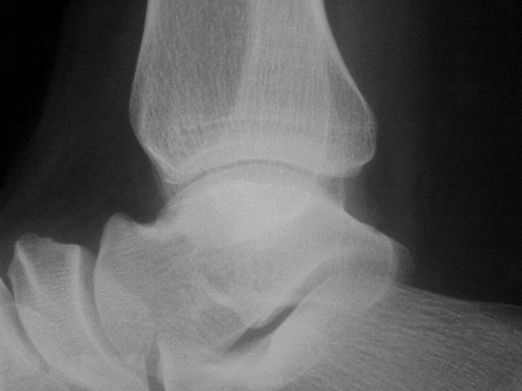 Initial study
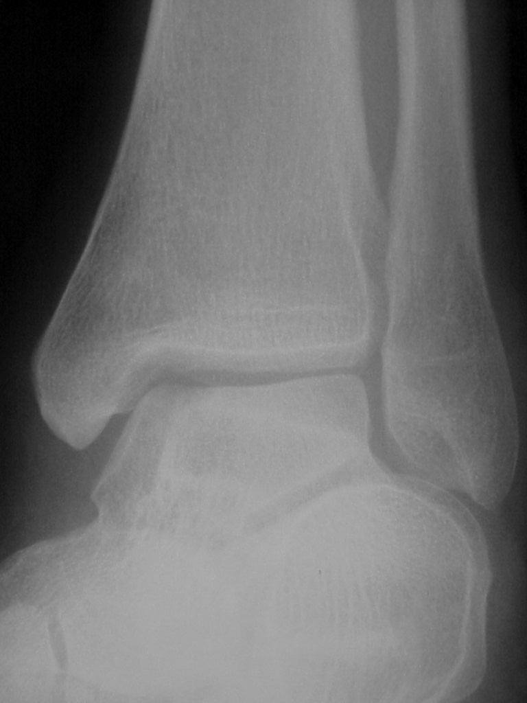
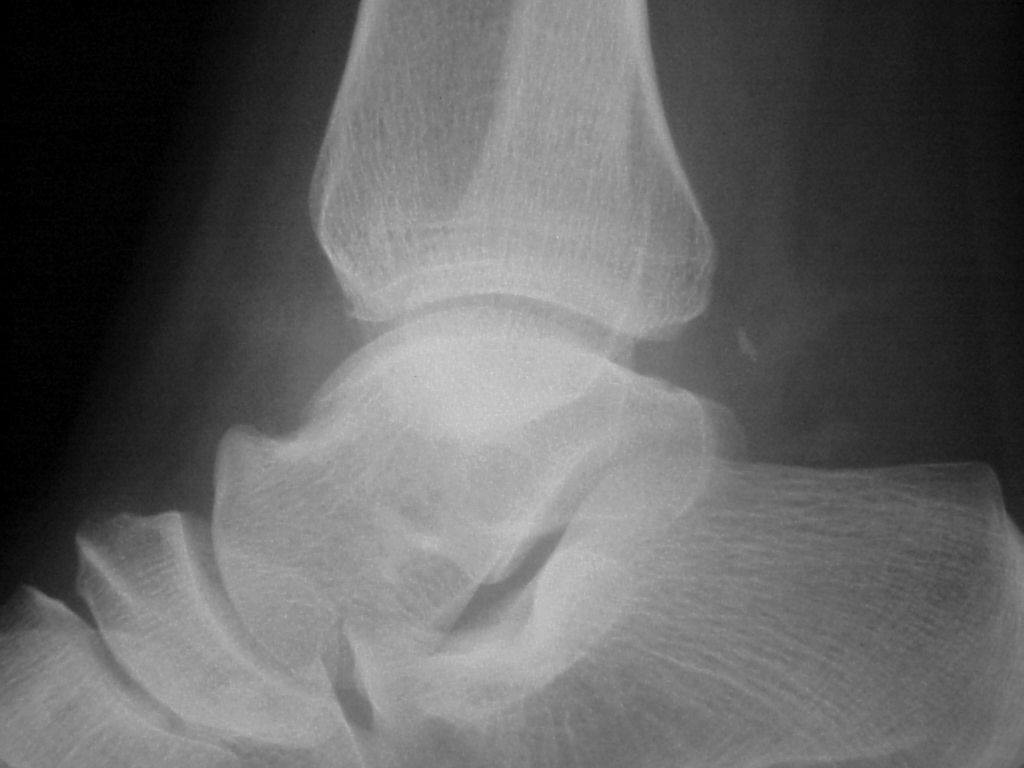 3 months later
Questions.
Describe the appearances and possible diagnoses. What has changed.
Answers.
The medial talar dome osteochondral fracture has displaced into the posterior recess of the joint.
Discussion.
Signs of a loose body rather than just an intraarticular body include movement with time. MRI can be useful to assess talar dome osteochondral injuries for loosening. T2 may show bright signal between the fragment and body of talus, but this is not diagnostic, since can also be caused by granulation tissue. Intraarticualr Gd MRI is the gold standard to assess stability. T1FS images before the injection of Gd are useful to ensure the high signal is due to Gd.